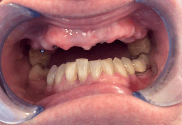
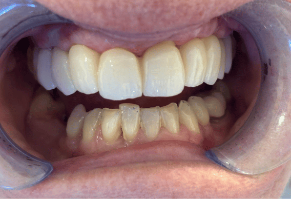
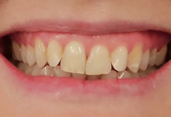
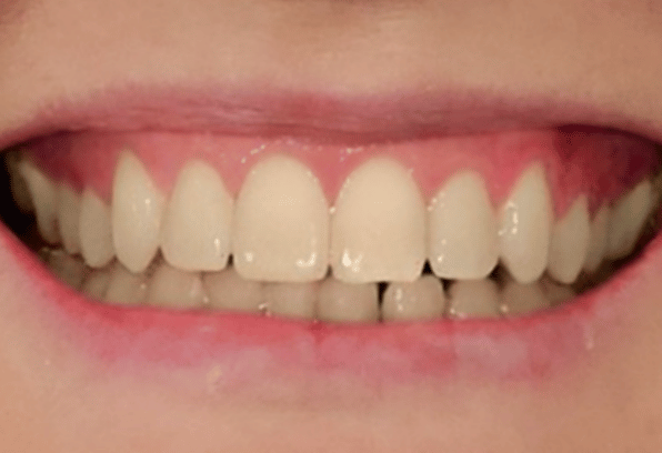
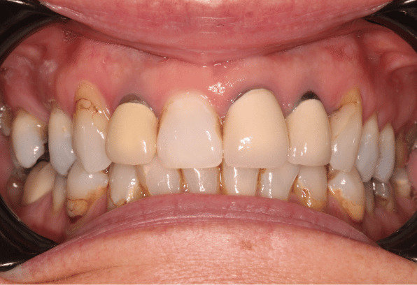
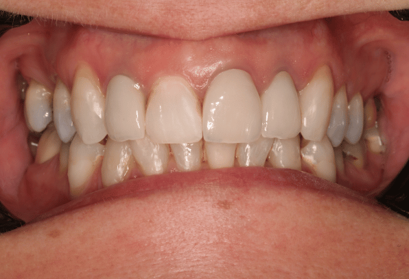
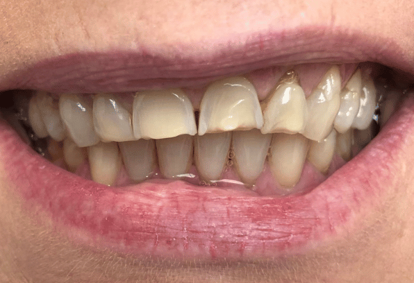
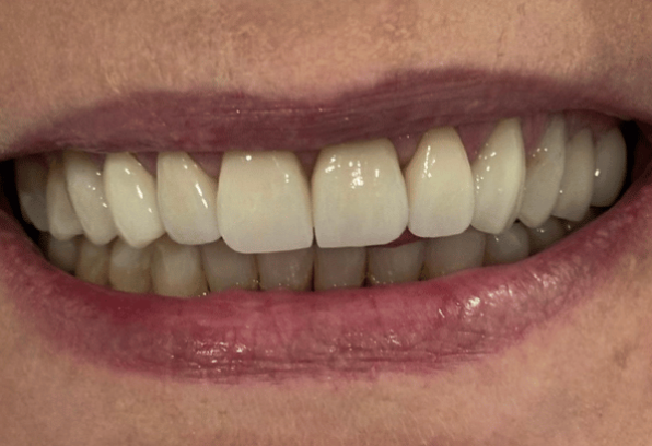
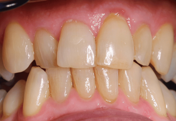
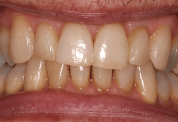
Patients without sufficient bone in their jaws to support implants can have an additional procedure to increase bone mass called a bone graft or block graft.
Once a tooth is lost or a tooth is extracted, the bone that once surrounded the tooth recesses or is lost because of the absence of the tooth.
Bone grafts increase the width and height of the bone at the implant site (where the implant needs to be placed). The surgeon performs a bone graft by using the bone graft tissue or material and attaching it to the deficient bone area.
The bone graft material contains collagen and proteins that also encourage bone growth. There are different types of bone grafts and different types of procedures performed for the upper and lower jaws.
Different types of bone grafts
Autogenous
This is where the bone is taken from somewhere else in the body (donor site), such as the chin, back of the jaw or, in some cases, the hip. A donor site taken from the patient means that there is a decreased risk of the body rejecting the bone.
Allograft
An allograft is a donated bone graft from another human. These work exactly the same as autogenous grafts.
Xenograft
These are where the bone is taken from an animal to be used for bone grafts. Either Equine or Bovine is usually used.
Alloplasts
This is a synthetic bone substitute, which is chemically similar to mimic human bone. Alloplasts also promote new natural bone formation.
Different types of procedures
Sinus lift or Augmentation
If there is insufficient bone mass in the upper jaw, a dental surgeon may suggest a Sinus lift. A sinus lift is where the bone in the upper jaw, where your molars and premolars are, is elongated. A bone graft is inserted between the jaw and the maxillary sinuses, which are on either side of the nose. In order to create space for the bone graft, the sinus membrane has to be moved upwards or lifted
Onlay graft
This is where the transplanted tissue or bone graft is added or laid directly onto the surface of the recipient’s bone. The new piece of bone will slowly join to the underlying region, and when healed and mature, an implant can be placed in a more favourable position.
FAQs
1. How is bone mass evaluated?
To evaluate a patient’s bone mass, a patient will need a CT scan, which is a computerised tomography. This advanced X-ray facility shows 3-dimensional photos of bone mass.
A CT scan enables a dentist to see the density of bone, the height, your anatomical structure and more. This will enable your dentist to evaluate your suitability for dental implant treatment as well as any additional procedures you may need.
Back to Dental Implants


